
Background:
Plants and animals are always competing to survive. There are many organisms that can produce substances to combat infections and viruses. Finding these substances can be used to potentially be turned into medicine. It is a long and difficult process to extract, process, and test these samples. Technicians have to deal with the extracts and then puriify them enough in order for them to be used.
Objective\Purpose:
What plant materials contain active ingredients that that will inhibit the growth of bacteria?
Materials:
-balance, weigh boat, lab scoops
-inoculating loop, Ni/Cr wire
-reaction tubes and rack, 1.7 mL
-LB broth base
-petri dishes, 60x15 mm, sterile
-methanol, absolute
-media bottles, 250 mL
-E. coli JM109 (stock plate)
-pipet, 1 mL and pump
-sterilizer, autoclave
-plant specimen
-dry block heater/heat block
-water bath, 37° C, shaking
-mortar and pestle
-forceps, fine-tipped
-laminar flow hood and disinfectant
-pipet, 10 mL and pump
-ampicillin
-glasses, safety, plastic
-plastic funnels, short-stemmed
-glass spreader
-Bunsen burner and gas lighter
-filter paper disks, 5 mm diameter
-incubator over, 37°C
-beakers, 100 mL
-syringe, 10 mL and filter, 0.2 um
Procedure:
1. Prepare a nutrient or LB culture for the E. colu at least 24 hours in advance. Using sterile technique, add a colony of E. coli culture to the broth medium and incubate, shaking at 37 C for 24 hours.
2. Each lab group need 2 petri playes. Draw a "+" on each plate bottom to divide the pate into quadramts (four sections). Label the quadrants No. 1 through 4. Also, label the dish with your initials and the date.
3. Liquefy sterile LB agar in the microwave at 50% power. Using sterile technique, pour approximately 20 mL of sterile, liquid LB agar into each Petri plate. Let the agar solidify for 15 minutes. Let it dry for at least 24 hours.
4. Using a mortar and pestle, grind up 2 g of plant tissue (leaves or bark) with 10 mL of deionized water. Lwt it sit for 3 minutes. Filter and sample through an 11 cm filter paper funnel. Filter sterilize the filtered sample extract using a syringe filter, as demonstrated by the instructor. Collect with 1 mL of extract into a 1.7 mL microtube. Label the sample.
5. Repeat Step 4, but replace the water with methanol as as the extracting solvent. After the methanol extraction, place the 1.7 mL tube with the 1 mL of methanol extract in a 65 C heat block (caps open) for 24 hours or more, if necessary to evaporate the methanol. Reconstitute dry matter in the tube with 1 mL of deionized water.
6. For each of the other samples, repeat steps 4 and 5. Label all samples. There should be six tubes of samples.
7. Using sterile forceps (that have been flamed in alcohol) drop three filter paper disks into each tube of the filtered extract.
8. Prepare negative control disks, three each, of only methanol and only sterile distilled water.
9. Prepare six positive control disks of ampicillin solution.
10. Allow the disks sufficient time to soak up enough extract to be saturated (perhaps overnight).
11. Close the tubes. Store all samples at 4 C until ready to use.
12. Using a sterile pipet transfer 1 mL of the E. coli broth
(made at step 1) to the middle of each Petri dish. Sterilize a spreading loop (using alcohol and a flame) , and evenly spread the bacterial culture around the Petri plate. Quickly cover, and allow the culture to soak into the agar for at least 15 minutes.
13. Using sterile forceps carefully place one disk into the middle of each quadrant, about 2 cm from the outer edge of the Petri dish. Blot any access liquid before placing the disk on the Petri dish. Keep all the methanol-extracted samples on the same dish and all the water-extracted samples on the same dish.
14. Repeat step 13 twice so that you have three replicates of the methanol extraction and three replicates of the deionized water extractions.
15. Place one of the negative control disks, either sterile distilled water or methanol, in the center of the appropriate plate. Place a positive control disk with ampicillin in another quadrant of each plate.
16. You should end up with six Petri plates, each containing a negative control in the middle, a positive control , and three sample disks. Make sure you have recorded exactly which plant extracts and which solvent went into each quadrant.
17. Make sure the disks are adhering well to the surface of the agar. For incubation, invert the plates and incubate at 37 C for 24 to 48 hours.
18. After incubation, examine the plates with the plant extract disks for zones of inhibition. This is a clear area formed around the disk by the inhibitory action of a substance(s) in the plant material. Photograph or draw the plates, labeling any inhibition of bacterial growth.
19. Create a data table to collect and present data of all the replicates as well as the averages. include descriptions of the bacterial lawn around each disk. Measure and record the diameter and clarity of any cleared areas around the disks. Give quantitative measurements of your observations.
The Results:
Here are the results of the lab!
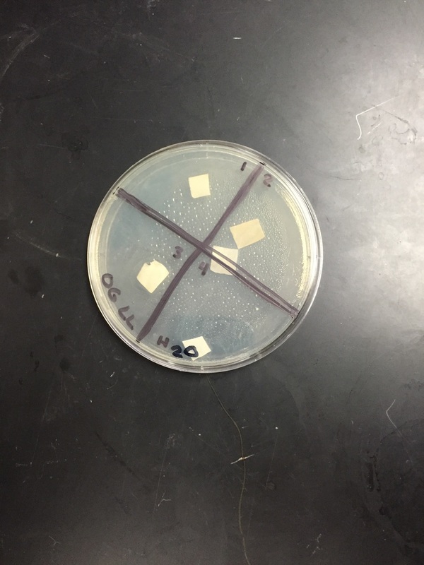
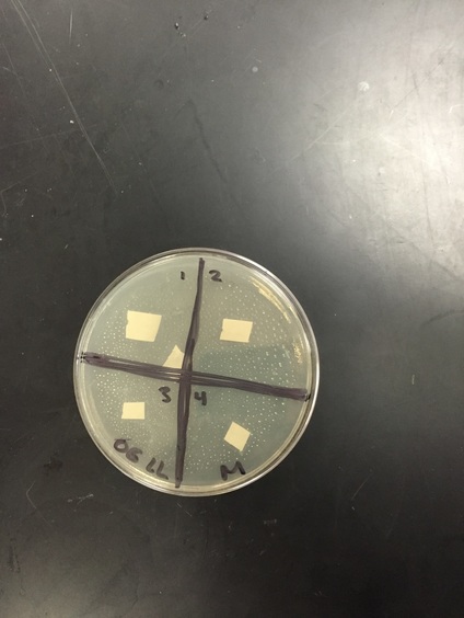
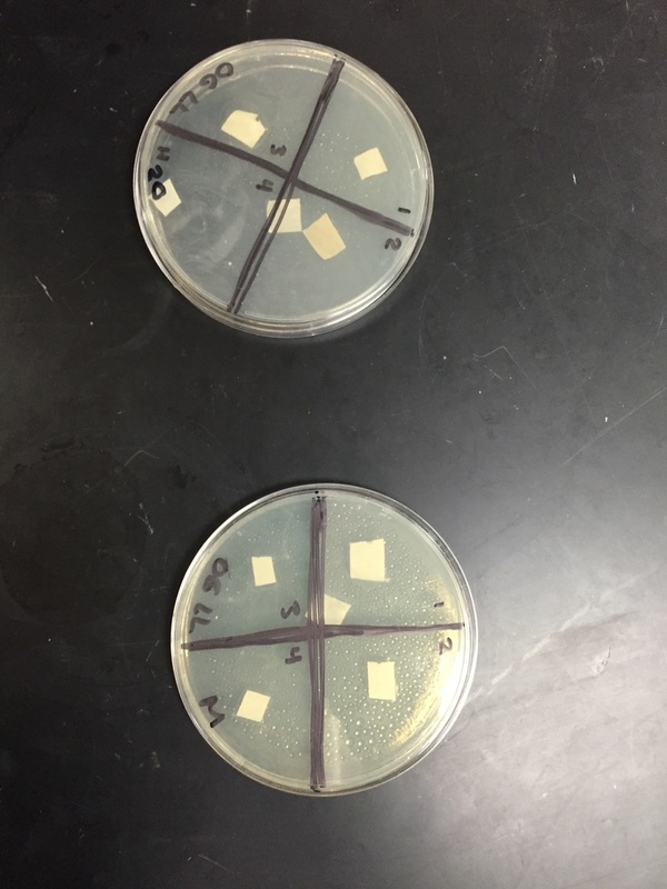
After 48 hours: There was an extremely small change with the slight ring of clear area around the paper in both the methanol dish and the water dish. The ring grew approximately 2 millimeters since yesterday. The bacterial lawn is more grown in, but still not totally complete. It looked like there was a slight contamination of our dishes on the first observation, but now it is gone.
Analysis/Conclusion:
There was a clearing around our positive control. We believe that we had a complete field of bacteria, but we aren’t positive. If we didn’t have a complete lawn of bacteria, the disk may not have absorbed enough of the ampicillin solution. The bacteria did grow around the negative control disks. Water is not expected to have a negative control disk or antimicrobial activity because water doesn’t kill bacteria, which means there is not any clearance around the paper. There was a very little clearance around our methanol plant extracts, and none that we could tell around the water plant extracts. Something that could have affected our results was a chance of contamination because of the bacteria on the desks or on our hands. Our plant could have antimicrobial activity, but we don’t know because we haven’t tested it. According to our results, lemon leaves don’t have any antimicrobial activity, because there was no or very little clearance around the extracts.
